16 June 2025
Ahmedabad Professors' AI Tool Challenges Biopsy to Target Brain Tumour
An AI-based digital solution for tumour characterisation has brought Ahmedabad University professors to the forefront of medical research. This development integrates advanced deep learning and radiomics to non-invasively diagnose and characterise brain tumours, addressing limitations in current diagnostic practices, which are heavily dependent on biopsies.
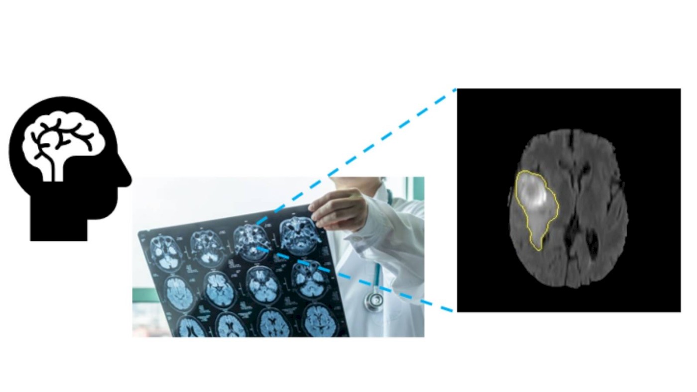
The challenge with current tumour diagnosis lies in understanding a tumour's microenvironment, which is crucial for accurately determining its grade and selecting the most effective treatment. At present, biopsies remain the gold standard for tumour diagnosis. However, they present several challenges: they are invasive, offer minimal information about the overall tumour, and can often be inconclusive or subjective during surgery. Such limitations may necessitate repeat surgeries, increasing patient burden and risk. This calls for a robust, non-invasive assessment procedure that can comprehensively examine tumour tissues using cutting-edge technology.
The research team includes Professor Jayendra M Bhalodiya (School of Engineering and Applied Science and the Bagchi School of Public Health) as the Project Investigator (PI), alongside Co-Investigators Professor Mehul Raval (School of Engineering and Applied Science) and Professor Kaumudi Joshipura (Dean and Professor at the Bagchi School of Public Health). The research team proposed an AI-based digital solution for tumour characterisation. The tool will be developed using convolutional neural networks (CNNs) and transformer-based architectures of deep learning alongside radiomics feature extraction methods. It will be tested using multi-sequence MRI data. Moreover, a feasibility study on glioma characterisation will be conducted to validate the tool using suitable histopathology and/or genomics data.
The team aims to develop a deep neural network-based tool that can process 3D volumes of multi-sequence MRI and provide segmented tumour areas for analysis using radiomics features. These features are quantitative measures that can be used to train a machine learning classifier to characterise tumours. Such AI tools can help determine tumour grade non-invasively and with greater accuracy. Furthermore, the derived radiomics features may help identify potential image biomarkers. Open-access data from the Cancer Genome Atlas (TCGA) repository will be used to develop the model and conduct experiments.
The development of this AI-based tool for brain tumour segmentation, characterisation, and diagnosis holds potential by offering a comprehensive, non-invasive, and accurate assessment method. It holds promise to revolutionise tumour diagnostics, potentially reducing the need for invasive biopsies and providing more precise information to guide patient treatment. This advancement could improve outcomes and enhance the quality of patient care.
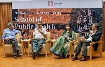
This research brings together experts from engineering and public health, reflecting a strong interdisciplinary approach to solving complex healthcare challenges. Supported by a University Challenge Grant, this project shows promise in steering a new pathway to precision medicine. Research at Ahmedabad University forms a central pillar of its academic vision, and through various initiatives, the University encourages faculty to engage with critical research questions. One such initiative is the University Challenge Grant, which is awarded to two or more faculty members from different disciplines to undertake interdisciplinary research that addresses complex societal challenges.